The lateral head radio graph has been a useful adjunct in orthodontics since its introduction in the 1930s. Over the years there has been a plethora of various systems of analysis, each with its own supporters. Before doing any analysis it is worth studying the film for its general proportions and relationships. The radiographs in Figs 1 and 2 are excellent examples. Both patients are in the 7 to 8 year-old age range. That said, the overall views show two very different individuals.
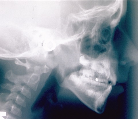
One of the most striking differences is the curvature of the cervical vertebrae. In Fig 1 the primary vertebral curvature has been lost, with the vertebrae resembling what chiropractors call a military spine. The transverse processes of C1 vertebra are touching the underside of the occiput when they should have at least 5-9 mm of space between them.
In Fig 2 the vertebrae show an excessive primary curvature, with large gaps opening anteriorly between the vertebrae. The space between the occiput and the transverse processes has opened up dramatically.
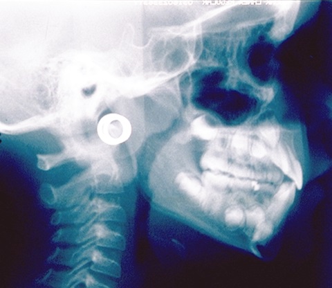
The cervical curvature is influenced to a large extent by the position of the occipital condyles. As they are positioned forwards or backwards they carry the C1 vertebra with them. The cervical curvature is therefore a useful indicator of what has happened to the position of the occiput. This in turn reflects the cranial shape, which has a compression of the cranium in the antero-posterior plane in Fig 1 and and an extention in the same plane in Fig 2. The osteopathic diagnosis was of a hyperflexion of the cranium in Fig 1 and a hyperextention in Fig 2.
There are other obvious anomalies (e.g., loss of airway in Fig 1 or the distal position of the maxilla in both cases). We will discuss these later. The point is that a careful study of the radiographs can yield all sorts of diagnostic clues even before any system of analysis is applied. The cervical curvature is therefore well worth such an examination.
Gavin

Leave a Reply
You must be logged in to post a comment.